Pet emergencies can be scary – we’re here to help you handle them.
We understand that seeing your pet sick, hurt or in distress can be stressful – especially if you don’t know what’s wrong. Fortunately, our experienced ER clinicians are prepared to take action and help.
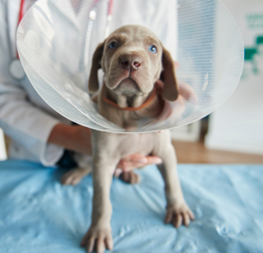
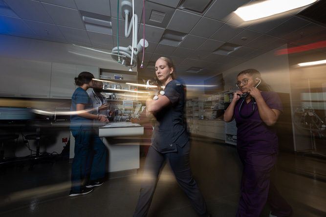
Acting quickly in an emergency may save your pet’s life.
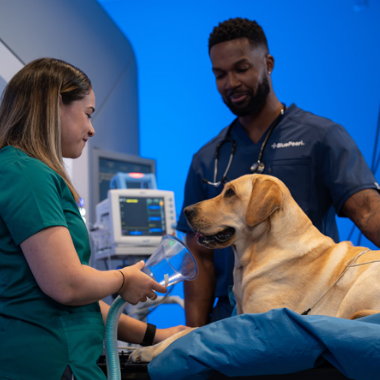
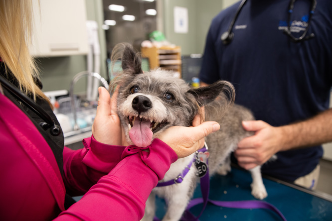
If your pet is experiencing an emergency, you’ll have an entire veterinary team helping them so you can get back to what matters most – spending precious moments together.
Experience makes all the difference.
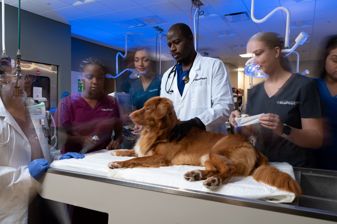
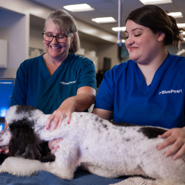
Our emergency team is made up of veterinarians, vet technicians and assistants, and support staff with rigorous training and experience in specialty medicine. The team works hand in hand to provide the comprehensive, compassionate care your pet needs and deserves. Because we’re a multidisciplinary hospital, the team can consult the expertise of other specialty departments, too.
Dr. Emily Ansel is excited by all aspects of emergency medicine. She especially enjoys cases involving internal medicine, toxins, wound care and reproduction.
Outside of work, Dr. Ansel enjoys reading, exploring the outdoors and spending time with her family. She has a special interest in purebred dogs and has participated in various dog sports with her Vizsla and Brittany dogs.
Dr. Katherine Blincoe brings a positive and bright attitude to work. She is particularly interested in emergency procedures including laceration repairs, thoracic limb amputations, foreign body removal, enucleations, and gastrointestinal (GI) surgery. She is also passionate about feline urethral obstruction and congestive heart failure cases.
Dr. Blincoe and her husband have a Labrador retriever, Wendy, and two cats, a black-and-white polydactyl, Rorschach, and an orange tabby, Shakespeare. She enjoys cooking, visiting with family, playing games, and spending time outside walking her dogs or drinking tea on her porch.
Dr. Lauren Block’s favorite cases are urgent emergencies. She loves stabilizing patients and seeing her efforts take effect. Her professional interests include urethral obstruction, pericardiocentesis, and hemoabdomen.
Dr. Block lives with her husband, young daughter and black Lab, Lacey. She enjoys exercising, staying active and traveling.
Dr. Kelly Catanzaro is interested in treating feline upper respiratory diseases as there are a myriad of potential treatments and she finds it gratifying to identify which ones work best for each patient. Her interests also include Lyme disease and other tick-borne diseases as well as infectious diseases, which were the subject of her PhD research.
Dr. Catanzaro and her husband have many pets, but her oldest (and most special) is her tortoiseshell cat named Hampster who she’s had since college. In her free time, Dr. Catanzaro enjoys hiking, being outdoors and renovating every room in her house.
Dr. Nick de Silvia’s professional interests include emergent endocrine diseases, autoimmune disorders and acute toxicities.
Dr. de Silvia lives with his wife, who is also a veterinarian, their two children, rescue mutt and two cats. Outside of work, he likes making homemade pasta and bread, exploring new places and playing pickleball.
As an experienced emergency clinician, Dr. Isel Del Valle is particularly interested in Eastern medicine, acupuncture, stabilization of septic patients, sports medicine, and rehabilitation.
Dr. Del Valle is passionate about travel, gourmet cooking, dressage, gardening, family time, and Duke University sports. She has a horse, a dog and a cat.
Dr. Tara Enzweiler appreciates the wide variety of cases, challenges, and fast pace of emergency medicine.
Dr. Enzweiler is an avid fan of Notre Dame Athletics. She enjoys spending time visiting North Carolina’s beautiful beaches, staying active, playing sports, and experimenting with new recipes in the kitchen.
A highly experienced clinician, Dr. Sarah Leigh is passionate about integrated, collaborative medicine. She thrives in the fast-paced ER environment and enjoys treating and managing complex cases.
A fan of outdoor sports, running and sports training, Dr. Leigh enjoys spending time with her husband, three children, dogs Stan and Shy and cat Joseph.
A highly experienced clinician, Dr. Kimberly Piner particularly enjoys treating cases of toxicity, urethral obstruction and parvovirus.
When not at work, Dr. Piner enjoys running obstacle races and spending time with her husband and two children. She has three cats, one three-legged calico, one bobtail and one polydactyl.
As an emergency clinician, Dr. Kelsey Routh communicates effectively with clients and uses essential problem-solving skills to diagnose and treat her patients. She finds cases involving shock stabilization, blood transfusions and internal medicine particularly gratifying.
Outside of work, Dr. Routh enjoys hiking, discovering new restaurants and spending time with her husband and German shorthaired pointer, Laney.
Dr. Rebecca “Becks” Schuber is most interested in diseases and disorders of the hepatobiliary system and endocrinopathies.
Dr. Schuber enjoys playing the piano, trivia and exploring the great outdoors. She has a fiancé, who is also a veterinarian, three dogs and a kitten.
Dr. Ashlan Westbrook is most interested in emergency and critical care cases, and she has previously worked in Ethiopia, researching epizootic lymphangitis in working equids.
Dr. Westbrook enjoys being outdoors with her golden retriever, Palmer, finding new music and bands, traveling, snowboarding and surfing.
Dr. Summer Wheeler’s special interests include wound care, treating cardiomyopathy and blocked cats, and performing ultrasounds.
Dr. Wheeler enjoys lifting, spending time with her fiancé, reading and watching movies. She also loves spending time with her cats, dogs and foster kittens.
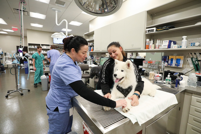
We know an unexpected trip to the emergency pet hospital can be stressful, and we want you to be prepared so you have one less thing to worry about. Our entire BluePearl team will be with you every step of the way.
During an emergency, every second counts. If your pet is experiencing an emergency, call us so we can talk you through their situation and be with you.
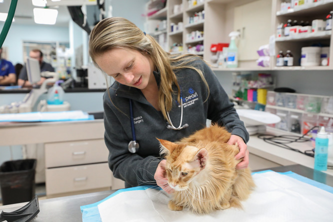
Our goal is always for your pet to have the best chance at a complete recovery so they can return home with you, happy and healthy.