Advanced cancer care to help your pet live a longer and better life.
We understand that seeing your pet experience unusual symptoms or act out of the ordinary can be stressful – especially if you don’t know what’s wrong. Fortunately, our veterinary oncology team is made up of experts in diagnosing and treating cancer, and we’re here to help.
Our commitment is not just to treat illnesses; it’s to enhance the well-being of your pet and be with you every step of the way.
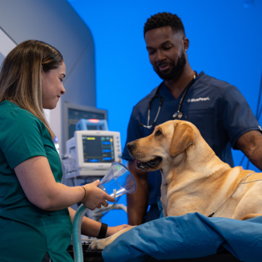
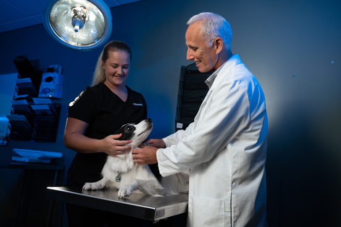
If your pet has cancer, you’ll have an entire oncology team helping to improve your pet’s quality of life so you can get back to what matters most – spending precious moments together.
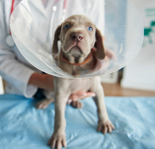
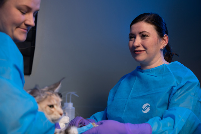
Your pet will receive unparalleled medical care during their time at the hospital (and they’ll be spoiled with love and attention, too).
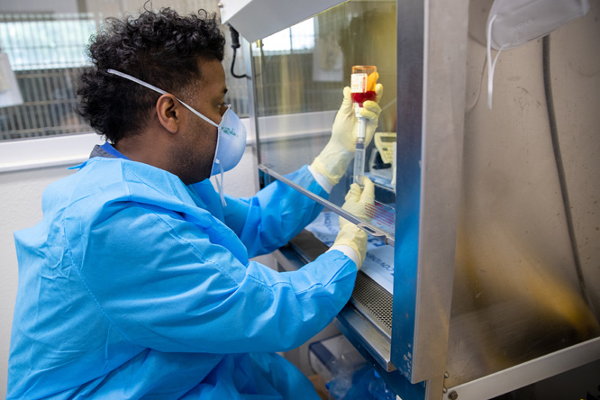
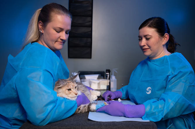
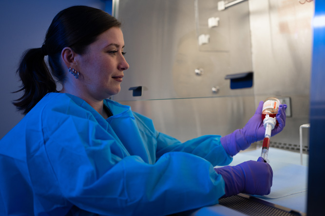
Your pet can’t tell us what’s wrong, so we use sophisticated diagnostics and imaging tools to uncover the source of the problem.
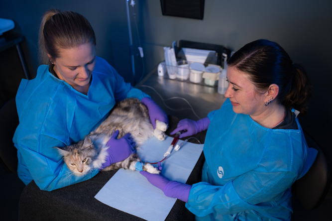
Just as no two patients are the same, neither are their treatment plans. Our oncology team has experience with a range of advanced procedures and minimally invasive options to get your pet on the path to wellness, including:
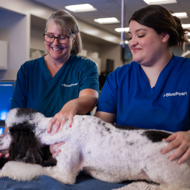
Experience makes all the difference.
Our oncology team is made up of veterinarians, vet technicians and assistants, and support staff with rigorous training and experience in specialty medicine. The team works hand in hand to provide the comprehensive, compassionate care your pet needs and deserves. Because we’re a multidisciplinary hospital, the team can consult the expertise of other specialty departments, too.
Dr. Rhiannon Doka provides compassionate and informative care for clients to make the best decisions for their pets’ cancer treatment options. Her goal is to give patients as much quality time as possible doing the things they love.
Dr. Doka’s special interests include comparative oncology, hematopoietic malignancy treatment, clinical trials, targeted cancer therapy and immunotherapy.
Dr. Doka enjoys spending her free time with family and friends. She has two dogs, Duffy and Taygan, and one cat, Rizzo, that always keep her on her toes.
Dr. Karri Miller obtained a master’s degree in veterinary science with a focus on oncology and currently collaborates on veterinary oncology projects with NCSU and Sentinel Biomedical. Her clinical interests include lymphoid cancers, cytology and surgical oncology principles.
Dr. Miller loves watching sports, especially college football, and supporting her Gators. She also enjoys spending time with her husband and daughter, as well as her three dogs, two Cavaliers and a Dachshund.
Dr. David Ruslander is interested in multimodality management for veterinary oncology patients, meaning treatment that involves surgery, radiation therapy, chemotherapy and/or immunotherapy to achieve the best outcome and quality of life.
Dr. Ruslander enjoys spending time with his wife, three children, two cats, dog and parrot.
Dr. Susan Shapiro is passionate about delivering personalized medicine and comforting end-of-life care; her favorite part of being a vet is getting to know pets and their families and helping them through difficult times.
Dr. Shapiro’s special interests include urothelial carcinoma in dogs, cancer genetics, cytology and molecular aspects of cancer, particularly how environmental exposures may relate to disease.
Dr. Shapiro lives with her husband, son and their dog, a sheltie named Gibita, who happens to be a cancer patient herself. In her free time, Dr. Shapiro enjoys singing, biking, running, international travel, good food and drinks.
Dr. Carly Stevens has worked in both a cell biology and genetics lab. Her clinical interests include treating histiocytic sarcoma, lymphoma, hemangiosarcoma and tumors.
When not working, Dr. Stevens enjoys bicycling, hiking, listening to podcasts, trying new recipes and watching movies. She has a mixed-breed dog adopted in California and named after a local beach.
We want you to be prepared for your pet’s visit to the oncologist, so you have one less thing to worry about. Our entire BluePearl team will be with you every step of the way.
Take the first step to getting your pet back to their best self – call us to speak with the oncology team and make an appointment.
Prioritize your pet’s health with the experts in specialty medicine.