Precision care for what matters most – helping your pet live a longer and better life.
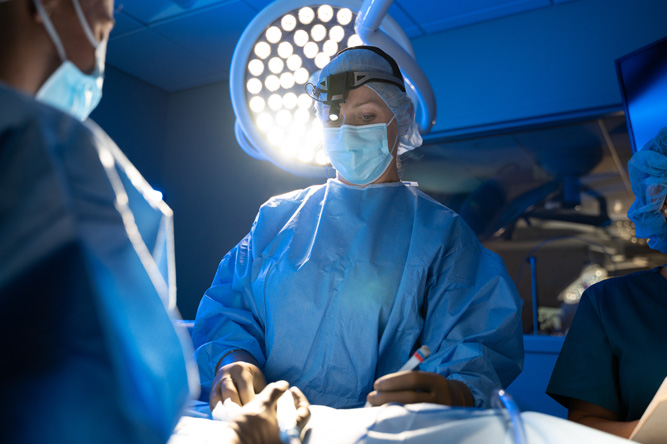
We understand that seeing your pet experience an emergency or condition that requires surgery can be stressful. Fortunately, our veterinary surgery team is made up of experts in performing complex procedures using the latest state-of-the-art techniques and equipment, and we’re here to help.
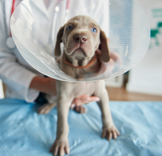
Our commitment is not just to delivering highly advanced surgical care; it’s to enhance the well-being of your pet and be with you every step of the way.
If your pet has a condition or injury that requires advanced care and procedures, you’ll have an entire surgery team helping to improve your pet’s quality of life so you can get back to what matters most – spending precious moments together.
Your pet will receive unparalleled medical care during their time at the hospital (and they’ll be spoiled with love and attention, too).
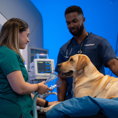
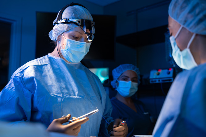
Your pet can’t tell us what’s wrong, so we use sophisticated diagnostics and imaging tools to uncover the source of the problem.
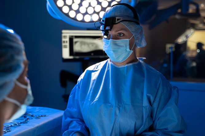
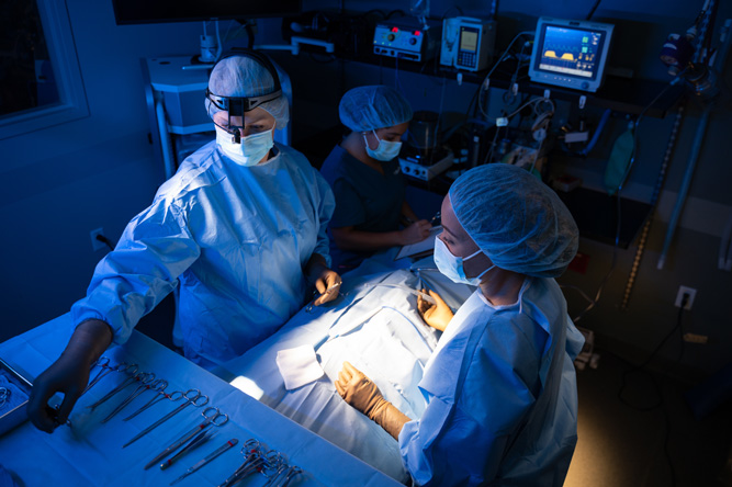
Just as no two patients are the same, neither are their treatment plans. Our surgery team has experience with a range of advanced procedures and minimally invasive options to get your pet on the path to wellness, including:
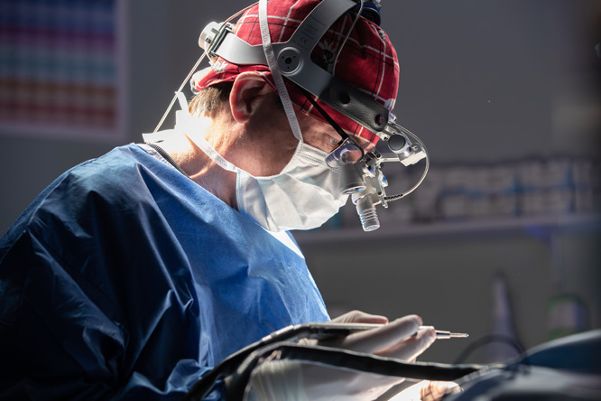

Experience makes all the difference.
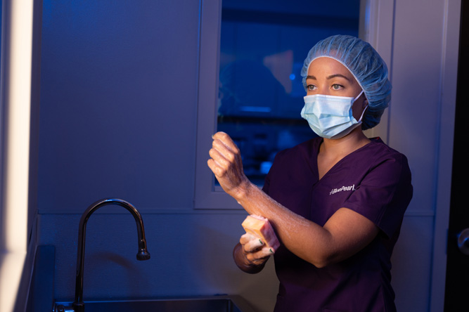
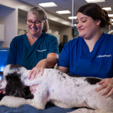
Our surgery team is made up of veterinarians, vet technicians and assistants, and support staff with rigorous training and experienced in specialty medicine. The team works hand in hand to provide the comprehensive, compassionate care your pet needs and deserves. Because we’re a multidisciplinary hospital, the team can consult the expertise of other specialty departments, too.
Dr. David Lee is most interested in orthopedic surgery, particularly surgical stabilization of the knee following cranial cruciate ligament rupture and joint replacement.
Dr. Lee enjoys woodworking, working about the house and watching his two sons play ultimate frisbee.
Dr. Marina Manashirova’s interests include surgical oncology, wound management, and any condition involving the thorax.
Dr. Manashirova’s family consists of her husband, daughter, and a 24-year-old ball python. She enjoys hiking, kayaking, and spending time with family and friends.
We want you to be prepared for your pet’s visit to the surgeon, so you have one less thing to worry about. Our entire BluePearl team will be with you every step of the way.
Take the first step to getting your pet back to their best self – call us to speak with the surgery team and make an appointment.
While a pet’s health problem is nothing to ignore, appropriate surgery and follow up will allow your pet to live a better and longer life.
Prioritize your pet’s health with the experts in specialty medicine.