Feline lower urinary tract disease: Signs, diagnosis and treatment.
Feline lower urinary tract disease will often only produce signs that mimic bladder infections. The bladder collects urine from the kidneys and contracts to expel its contents during urination via a tube called the urethra. The end of the urethra in a normal cat is relatively narrow. Early neutering of male cats may cause the urethra to fail to develop to a normal diameter.
In some of the affected cats, mucous plugs, crystals and stones can become lodged in the terminal part of the urethra of a male cat, which results in urinary obstruction. Over a period of 24 hours of obstruction, toxins build up in the cat’s body and can cause death.
Signs and symptoms.
Shortly after urinary obstruction occurs, the affected cat will go to the litter box and attempt to urinate without success. As the 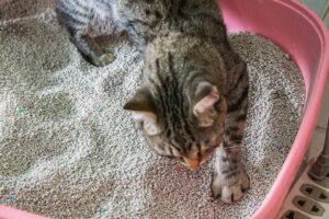 bladder becomes more and more distended, signs of pain may be evident: the cat may seclude himself, cry out or growl.
Upon palpation of the abdomen, the bladder will be painful, firm and the size of an orange. As the toxins accumulate in the blood, the patient develops signs of vomiting, abnormal heartbeats, depression, and in severe cases coma. These toxins can be fatal to the patient.
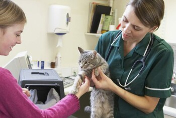
Diagnosis.
Finding a very large and firm bladder upon physical examination makes the diagnosis of urinary obstruction most probable. X-rays of the abdomen are necessary to rule out obstruction caused by small stones in the urethra and bladder. Blood is tested to evaluate for accumulation of urinary toxins and high potassium levels. Abdominal ultrasound can also be used to evaluate the urinary tract but may not be necessary in every patient.
Treatment.
Urinary blockage is an emergency; therefore, it is essential to have this condition treated immediately. Your veterinarian will start intravenous fluid therapy, as these patients commonly are dehydrated. If the potassium level is very high, glucose (a type of sugar) and insulin are administered intravenously to lower the level of this electrolyte. The urethral obstruction is relieved by flushing the material out of the urethra and passing a urinary catheter into the bladder. If present, stones are surgically removed from the bladder once the patient is stable and the toxins have been eliminated from the bloodstream.
Most patients will have a urinary catheter left in place for at least 24 hours after the obstruction has been relieved. After the catheter is removed, the patient is carefully monitored for urination. In the event that the patient becomes obstructed again, the obstruction will need to be relieved again.
Perineal urethrostomy.
If the patient becomes repeatedly blocked, a perineal urethrostomy is recommended. The perineal urethrostomy essentially removes the narrow terminal portion of the urethra, which is located within the penis. During the procedure, the urethra is cut open approximately 0.5 inch, to a point where the urethra enlarges. The edges of the urethra are then sutured to the edges of the skin using very fine sutures.
For more information on this subject, speak to the veterinarian who is treating your pet.