Radiology revealed.
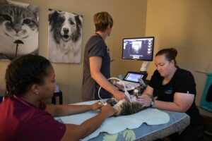
Clients often ask me, “How is an MRI different from a CT?” or “If my vet already did x-rays, why does my pet need an ultrasound?” These are very good questions, as each type of imaging works differently, shows different parts of the body and of course have a wide range of cost associated with them.
Imaging technology as a diagnostic tool.
 Radiology is a branch of medicine that uses imaging technology to diagnose disease. Radiology is a crucial element in diagnosing and treating patients in veterinary medicine. Imaging devices may use radiation, pulses of high-frequency sound or magnets to create images of specific areas of a patient where this disease is suspected. Are we looking at a patient’s brain, heart, bones, spinal cord or an abdominal organ? This tells us what imaging device to use in order to find what we’re looking for. Those images can then be used to determine the best treatment options for that patient.
Images of some diseases are more complicated than others and require the interpretation of a board-certified veterinary radiologist, who has received advanced training in diagnostic imaging and passed the American College of Veterinary Radiology Examination.
Radiographs “x-rays.”
Radiographs are images produced on a sensitive plate or film by x-rays, gamma rays, or similar radiation. They are used as a good starting point for orthopedic diseases, such as bone fractures, and a method to rule out abdominal concerns like foreign bodies and bloat. They provide a two-dimensional picture and not all structures are equally visible. For example, the spinal cord cannot be seen on radiographs, although we can see vertebral 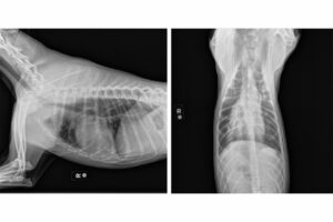 bodies and disc spaces. They cannot be used as a diagnostic tool for intervertebral disc disease or to diagnose a herniated disc.
Some orthopedic changes can be seen on radiographs, but still require additional advanced imaging to diagnose. Radiographs like the examples below, can be used to find thoracic disease including metastatic cancer prior to an anesthetic procedure. We can see metastatic changes as well as primary lung tumor changes, asthma, pulmonary edema and other pulmonary conditions. Radiographs can usually be taken with the patient awake, however for good positioning in certain conditions, sedation may be needed.
Ultrasound.
Ultrasound uses high-frequency sound pulses to diagnose a wide range of diseases including cardiac, abdominal, ocular and thyroid diseases. Anything surrounded by bone and air cannot be visualized with ultrasound. It shows us structures in real-time, giving us a multi-dimensional view of organs like the heart, liver, kidneys and pancreas.
Below is the ultrasound of a cat’s kidney. The visual structure of this kidney gives us information about the health of this organ. Although radiographs are a good starting point, they may not reveal architectural abnormalities in less dense structures like these. Ultrasound can be performed without anesthesia, although sedation is sometimes needed for anxious patients.
CT (computerized tomography).
A computerized tomography (CT) scan combines a series of x-ray images taken from different angles around the 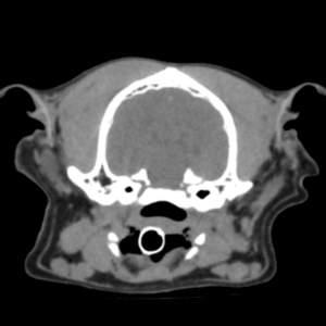 patient’s body and uses computer processing to create cross-sectional images (slices) of the bones, blood vessels and soft tissues. CT scan images provide more detailed information than x-rays.
CT scans can be used to locate such pathologies as primary tumors and metastasis in the thorax and abdomen, abnormalities in the nasal cavity and ears when bulla disease is highly suspected, joint conditions and long-bone complex fractures. It is a multi-dimensional imaging modality and can usually be performed with sedation but may require anesthesia in certain situations.
MRI (magnetic resonance imaging).
Magnetic resonance imaging (MRI) is a medical imaging technique used in radiology to form pictures of the anatomy and the physiological processes of the body. MRI scanners use strong magnetic fields, magnetic field gradients, and radio waves to generate images of the organs in the body. MRI is primarily used for imaging the brain, spine and spinal cord, however regions such as the shoulder, eye, and stifle are becoming more popular (surgeon dependent).
MRI shows details in the nervous system that cannot be seen on CT or radiographs. We can view three-dimensional images of the area being scanned. Anesthesia is required because patients need to be immobilized for image quality and due to the length of the scan which is typically over an hour.
Side by side.
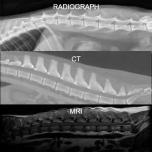
This example shows three imaging methods of a canine spine, side-by-side to emphasize the differences. The spine is made up of three parts; the vertebrae, the intervertebral discs and the spinal cord. The top image is a radiograph. This method reveals clear definition of each vertebra and can be a good way to find a fracture or other changes in the bone.
The middle image is a CT. This method clearly shows spacing between each of the vertebrae, clearer bone definition and bone density. The bottom image is an MRI. With this method, we can see the spinal cord within the vertebrae. This is used to diagnose neurologic disease.
 In conclusion, whatever the diagnosis, we have the tools to find it, and then do our best to treat it. Thinking about medical advances, it is astonishing that we have these capabilities for our pets.