Feline Fibrosarcoma: Signs, Treatments and Results
In 1985, two feline aluminum adjuvant vaccines, feline leukemia and rabies, were developed and subsequently used clinically. A few years later, veterinarians were reporting vaccination reactions.
Some of these were biopsied and found to have inflammation with a core of brown unidentifiable material. It was suspected that the material was the aluminum adjuvant, the vaccine itself or a combination thereof.
In 1991, Hendrick reported the first vaccine-associated sarcoma. Others have reported the development of a sarcoma at the site of the excised vaccination reaction. Some cats developed multiple sarcomas located at each vaccination injection site. Epidemiologic associations first were made with feline leukemia and rabies vaccination and later with feline panleukopenia and feline rhinotracheitis vaccines.
Vaccine Reactions
Cats can develop a lump (mass) at the site of a vaccination injection. Usually the mass will resolve spontaneously and does not form into a cancer. In general 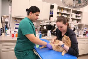 one in 10,000 vaccinated cats will develop cancer due to the vaccination. If a mass 1) has been present for more than three months, 2) is greater than 2 cm in size, or 3) has increased in size one month after the vaccination was administered, then a biopsy of the mass should be performed.
If a cat has had a tumor removed due to vaccinations and later is vaccinated, early tumor recurrence is likely. Vaccine-associated sarcomas can develop in the layer of connective tissue between the skin and muscle. These tumors have microscopic cells that extend like roots deeply into surrounding tissues. For this reason, recurrence of the tumor following tumor surgical removal is very common.
At the time of diagnosis, only 3-5% of the tumors have spread (metastasized) to the lungs, skin, subcutaneous tissues, region lymph nodes, chest, liver or pelvis; however, with time the spread rate increases to 24%.
Signs and Diagnosis
The typical sign of a vaccine-associated sarcoma is a mass (lump) at the site of a vaccine injection site. The most common locations for vaccination-associated masses include the back, shoulder blades or on a limb, as these correspond to vaccination injection sites. The mass commonly is firm, non-painful and often attached to the skin or underlying muscles.
Most patients do not feel ill when the lump is first found. If left untreated, these masses can become ulcerated and infected. With time, if they spread to the lungs and other parts of the body, they can cause breathing difficulty, anorexia and weight loss.
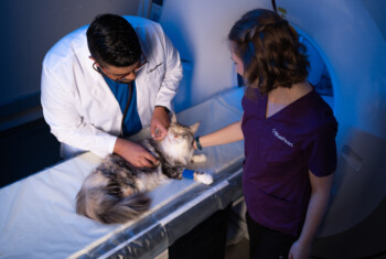
The diagnosis of a vaccine-associated sarcoma necessitates a biopsy. Additional tests performed prior to surgery may include a complete blood count, blood chemistry profile, feline immunodeficiency virus infection test, feline leukemia virus test, and urinalysis to check internal organ health. Chest X-rays and abdominal ultrasound are used to identify visible spread of the cancer; however, microscopic spread of the tumor to other organs cannot be detected with X-rays or ultrasound. A CT scan helps the surgeon develop a plan to surgically remove the tumor.
Treatments
Surgery is an essential treatment for vaccine-associated sarcomas. For the best outcome, the tumor and a large zone (2 to 5 cm) of surrounding “normal” 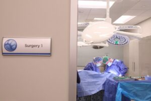 skin, fat and muscle should be removed. If the tumor is located on a limb, amputation is essential for a good outcome.
Chemotherapy may be recommended and is administered every three weeks via intravenous injection by an oncologist for a total of four to five treatments. The treatments are typically done on an outpatient basis and may take a total of 90 minutes to complete each visit. Unlike humans, most cats do not lose their hair and usually have only mild side effects from the medication such as transient loss of appetite and vomiting.
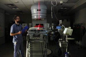
Radiation therapy has been shown to improve the survival of patients that have had surgery and may be used to shrink an inoperable tumor or used to kill residual cells that may be present. Radiation is administered daily until 18 to 21 treatments have been completed.
Results
If the margins of the surgically removed tumor are not cancer free and no further treatment is performed, the median disease free interval (time until tumor recurs) is reported to be two to six months. With clean margins and no additional treatment, median disease free interval is 276 days and median survival time is 576 days.
 With clean surgical margins and radiation therapy, the disease free interval is one year with a median survival time of two years. In another study, preoperative radiation followed by surgery resulted in a disease free interval of 986 days with clean excision of the tumor versus 292 days with incomplete excision. A more recent report indicated that surgery and radiation with clean surgical margins had a disease free interval of 37 months and median survival time of 43 months.
Short-term complications following surgery are uncommon and may include temporary dehiscence (opening) of the incision and infection. Tumor recurrence and spread of cancer are other complications. Side effects of radiation may include skin burns, poor healing of the incision, dermatitis and regrowth of hair coat that is a different color (grey or white).
For more information on this subject, speak to the veterinarian who is treating your pet.