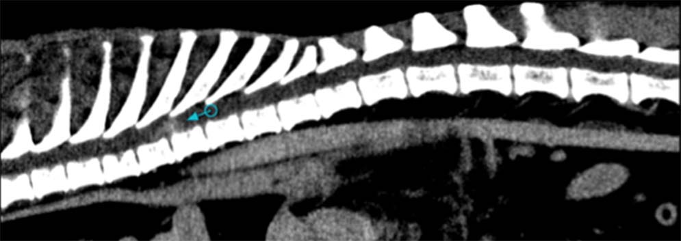
Figure 5. A poorly circumscribed, contrast-enhancing intramedullary mass is noted within the midthoracic spinal cord on CT (arrow). No definitive diagnosis was made, but neoplasia is suspected in this geriatric canine patient.


Figure 5. A poorly circumscribed, contrast-enhancing intramedullary mass is noted within the midthoracic spinal cord on CT (arrow). No definitive diagnosis was made, but neoplasia is suspected in this geriatric canine patient.