Intestinal intussusception: Signs, diagnosis and treatment.
An intussusception causes an obstruction of the bowel and can compromise its blood. It is the invagination or telescoping of one part of the bowel into an adjacent part of the bowel.
Anatomy.
The gastrointestinal tract consists of a tube that runs from the mouth to the anus. Its function is to digest food and absorb nutrients into the body. The stomach is a dilated part of the GI tract that produces acid, which helps with the initial breakdown of proteins. The small intestine extends from the stomach to the colon and serves to further break down food into absorbable nutrients. The colon is the reservoir for stool, serves as a water absorber, and is the site for production and absorption of certain vitamins.
Intussusception.
This is similar to collapsing a telescope together. An intussusception is incited by abnormal motility (movement) of the bowel which can be caused by viral infections, bacterial infections, intestinal parasites, intestinal foreign bodies, dietary changes, intestinal tumors, and surgical procedures previously performed on the bowel.
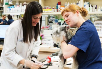
Signs and diagnosis.
The primary signs of intussusception include vomiting, diarrhea, anorexia, depression and dehydration (mouth becomes less  moist and saliva becomes tacky). Crying, whimpering, unwillingness to lie down, or assuming a hunched or praying position (down on the forelimbs and up on the hind limbs) may be a sign of abdominal pain. Some dogs afflicted with an intussusception located at the terminal part of the small intestine may have signs for weeks including progressive weight loss, vomiting and poor appetite. In other cases, the intussusception may protrude out of the anus.
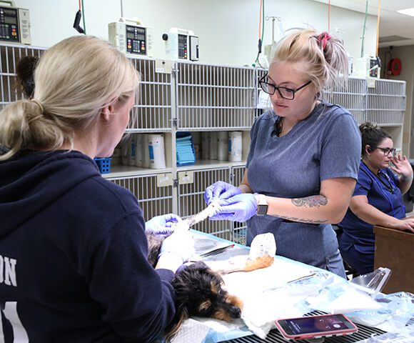
The diagnosis of an intestinal intussusception generally is made with a physical examination (abdominal palpation) and x-rays. Abdominal ultrasound is an excellent tool to diagnose intussusception. Blood work is used to evaluate overall health of your companion and to help the veterinarian direct stabilizing treatments prior to surgery. The veterinarian will tailor a diagnostic plan for your companion that will allow for an expedient diagnosis and treatment.
Treatment.
An incision will be made into the abdomen to allow the surgeon to examine the internal organs. If the bowel affected by the intussusception is in good condition, it is massaged to separate the telescoped bowel. In most cases, the affected portion of bowel 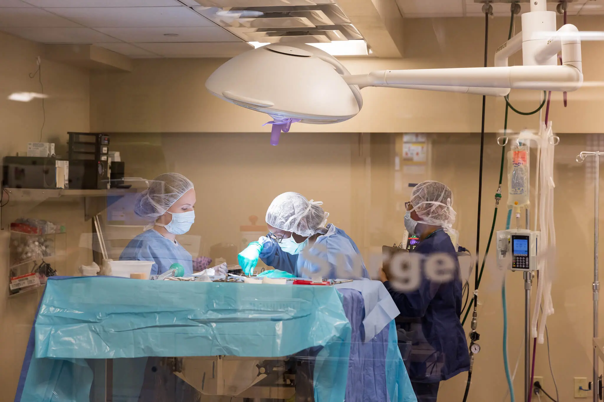 must be removed and the bowel surgically reconnected.
Up to 20% of patients that are surgically treated for an intussusception may have a recurrence of the problem; however, if adjacent loops of bowel are tacked to each other with sutures (called “surgical plication”) this problem can be prevented.
Occasionally, an intussusception can spontaneously reduce by itself and the bowel will appear normal at the time of surgery; however, the intussusception commonly recurs if the bowel is not surgically plicated.
While in our hospital, your companion will continue to receive intravenous fluids, electrolytes and in some cases plasma or an artificial plasma product called hetastarch. Your companion will be carefully monitored in the intensive care and will be given narcotics to ensure a pain-free recovery. Most patients that have abdominal surgery leave our hospital within 48 to 72 hours.
For more information on this subject, speak to the veterinarian who is treating your pet.