What are brain tumors?
Brain tumors are a type of cancer that occurs in the brains of dogs and cats. Like many areas of the body, cancerous cells can grow and create a tumor. However, with brain tumors, there is no extra room for a tumor to grow within the skull, so they can crowd and cause pressure within the brain. This can lead to problems from the tumor itself, but also from the pressure caused by the tumor.
Clinical signs and symptoms
With neurological problems, the clinical signs or symptoms are related to the location within the brain, rather than the type of disease. This is why there is a lot of overlap in symptoms between various brain diseases.
Tumors located in the forebrain (the front part of the brain) of dogs and cats commonly cause:
- Behavior changes
- Confusion
- Agitation
- Seizures
Tumors located in the brain stem (the back part of the brain) commonly cause:
- Balance issues like vertigo
- Incoordination and weakness in the limbs
Tumors located in the cerebellum commonly cause:
- Tremors
- Balance issues
The pressure created when a tumor crowds the brain can also cause mental dullness, or can exacerbate any of the other symptoms listed above.
Causes
Brain tumors are typically named after the type of cell it comes from. Brain tumors can arise from within the brain cells in the tissue of the brain (such as gliomas, astrocytomas or oligodendrogliomas), or they can arise from the meningeal lining of the brain (such as meningiomas).
Other types of brain tumors include:
- Choroid plexus tumors
- Lymphoma
- Pituitary tumors
- Histiocytic tumors
Less commonly, a tumor may arise from the skull and bones (e.g., osteosarcoma or multi-lobular tumor of the bone).
In all cases, tumors arise when cells start dividing uncontrollably.
Benign and malignant tumors
Some brain tumors are “benign,” meaning they do not tend to spread to other organs of the body. Other tumors are “malignant,” meaning they can spread to other organs. This is called metastasis.
Metastasis means the tumor cells can get into the blood or lymphatic systems of the body and spread to other organs. Common sites of metastasis are the lungs, liver, spleen and bones, although metastasis can affect any part of the body.
Similarly, tumors from other parts of the body can spread to the brain as well. For this reason, your veterinarian may recommend screening other organ systems to “stage” the cancer, which determines if any other organs are affected.
Regardless of whether a brain tumor is benign or malignant, they all can have a serious impact on the patient. Both benign and malignant brain tumors require treatment because there is not enough room in the skull for a tumor to grow without causing damage to the brain.
Getting a diagnosis
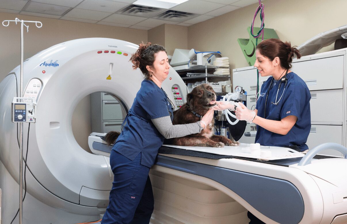
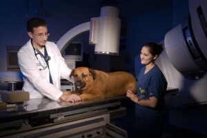
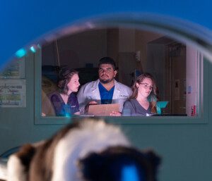 MRI (magnetic resonance imaging)
Because the nervous system is so hidden away from view, an MRI scan is required to diagnose a brain tumor. This allows your veterinarian to see the tissues of the brain in great detail.
An MRI scan takes about an hour to perform. During that time, your vet will use different settings to highlight various characteristics of the brain tissue. Some settings are better for showing tissue while others show fluid, inflammation, hemorrhage or ischemia (e.g., strokes).
These images are captured again after the patient is given an intravenous contrast agent. Contrast agents show up bright white on the MRI scan and are used to highlight tumors and inflammation.
Spinal fluid analysis (spinal tap, CSF tap)
In some cases, a spinal tap to evaluate spinal fluid may also be important for achieving a diagnosis. An MRI is often performed first because it may provide a diagnosis on its own and can help determine if a spinal tap is safe to perform. In many cases, if a tumor is present, this causes pressure within the brain. Performing a spinal tap when there is increased pressure within the brain may be very dangerous and could even be fatal.
What does an MRI reveal?
An MRI can help determine the location and size of a tumor, whether it is crowding the normal brain tissue, and tissue characteristics that indicate what tumor type the mass may be. This is important as different tumor types have different prognoses.
What are the limitations of MRI?
An MRI provides a lot of helpful information; however, it is not always perfect. It can often determine the likely tumor type, but a diagnosis is never certain without a biopsy. A biopsy allows a veterinarian to look at the tumor cells under a microscope and determine the specific tumor type with great accuracy. This is called a histopathologic diagnosis and is the most accurate form of diagnosis available.
How is a biopsy performed?
There are two types of biopsy procedures:
- Minimally invasive (e.g., needle biopsy)
- Surgical (e.g., an open biopsy)
Minimally invasive biopsies use different techniques to guide needle placement:
- Anatomic landmarks such as the appearance of skull shape
- Image guidance like a CT or MRI scan
- A neuronavigation system such as a stereotactic navigation computer system
With needle biopsies, a small burr hole is made in the skull to allow the needle to pass, using a surgical drill similar to a Dremel drill, and a small sample of cells is drawn into the needle and submitted for analysis.
For a surgical biopsy, the goal is to remove as much of the tumor as possible while also collecting cells for the microscopic evaluation. During a surgical biopsy, a small window is made in the skull to access the tumor. The tumor is gently removed using specialized instruments.
Questions for your doctor
When consulting with your pet’s veterinarian, consider asking questions like:
- What are the treatment options, risks and predicted outcomes for tumors of this type in this location?
- Are there any clinical trials available in our region for this type of tumor?
- Can you provide me with a referral to a specialist?
Treatment
The most common treatment options for brain tumors are surgical resection (removal) and radiation therapy.
Chemotherapy is not typically used for brain tumors, with rare exceptions, as many chemotherapy agents cannot cross the blood-brain barrier to reach the tumor and make an impact.
Surgical resection
Not all tumors are suited for surgical resection (also called surgical excision or surgical removal). In order to be surgically accessible (or reachable), the tumor must be near the surface of the brain. Deeper tumors are not typically accessible by surgery because the normal brain tissue overlying the tumor may be damaged in the process, although some neurosurgical centers have specialized equipment that allows safe surgical access to these deep tumors.
Surgical resection is beneficial in many ways:
- It allows for histopathological diagnosis, which is the most accurate means of diagnosing a brain tumor using a microscope to evaluate the cells within the tumor.
- It also serves as a treatment for removing as much of the tumor as is safely possible. Even if some of the tumor cannot be removed, surgery will alleviate some of the pressure within the brain by reducing the volume of the tumor (this is called “debulking”).
Although there is a benefit to performing surgery, it will never provide a “cure” for a patient with brain cancer. This is because it is never possible to remove all of the tumor cells. Microscopic cells may remain or visible tumor cells may not be able to be removed safely, and a portion of the tumor must be left behind.
Tumors on the skin, for example, can be removed along with a halo of normal tissue (usually 1 to 2 centimeters of normal tissue beyond the tumor) to ensure that all of the tumor cells are removed. This is not possible in the brain, because the normal tissue surrounding the tumor is essential to function. For this reason, only the tumor itself can be removed, and even then, sometimes a portion of the tumor cannot be removed safely.
 Radiation therapy
 Radiation therapy
Radiation therapy is a non-invasive treatment that can be used as a sole therapy or may be used in combination with surgery.
Radiation therapy delivers a dose of radiation to the tumor to damage the tumor cells and make them stop growing. This is similar to having an x-ray performed, only using a bit fancier technology. There are two types of radiation therapy available:
- Stereotactic radiation therapy (also called gamma knife, stereotactic radiosurgery, SRT or SRS)
- Fractionated radiation therapy (including intensity-modulated radiation therapy (IMRT))
Stereotactic radiation is delivered using high-dose treatments over a few sessions. Stereotactic radiation therapy is typically delivered on consecutive daily schedules or every other day for approximately 1 week.
Fractionated radiation therapy is delivered using low-dose treatments administered daily for approximately 20 sessions.
There are certain situations where stereotactic radiation therapy or fractionated radiation therapy may be preferred for a particular patient, tumor type or tumor location. A radiation oncologist can discuss the best treatment plan for your pet.
Prevention
Currently, there are no known methods for preventing brain tumors. Clinical trials are underway to evaluate whether a pre-administered vaccine can reduce the occurrence of cancer in dogs, but these results will not be available for quite some time.
Prognosis
The prognosis for brain tumors varies widely depending on the tumor type and tumor location.
Meningiomas
Meningiomas tend to be one of the more responsive tumors to treatments. The average survival of patients with meningiomas may range from 1-3 years, depending on tumor location, and whether surgery, radiation therapy or combination therapy is administered.
For patients treated only with supportive care to maintain comfort and control symptoms, the median survival may be less than 6 months.
Gliomas
Gliomas (e.g., gliomas, oligodendrogliomas and astrocytomas) tend to respond poorly to treatment. Patients with gliomas have an average survival of 1-2 months without treatment. With radiation therapy, average survival is typically 6 months. It is uncommon, but some patients may live quite a bit longer.
Many BluePearl Pet Hospitals around the country offer neurology and oncology specialty veterinary services. Find a BluePearl neurologist or oncologist near you.