Lung lobe tumors in dogs and cats: Signs, diagnosis and treatment.
The lungs have a number of functions including transferring oxygen from the air into the blood, transferring carbon dioxide from the blood to the air, metabolizing a number of drugs, and controlling blood pressure.
Blood from the entire body is pumped through the lungs many times each hour, which helps to filter out bacteria and cancer cells. If cancer cells actively divide into more and more cells, a tumor will develop in the lobes of the lungs. This is called a metastatic lung tumor as it is from another part of the body.
It is common that this type of tumor originates from one of the internal organs: mammary glands, the skin, connective tissues or bones. These tumors are typically spread throughout the lung fields; therefore, multiple masses are seen in the lungs on x-rays.
In contrast, the cells of lung tissue can transform into a cancer and grow into a mass. This type of tumor is called a primary lung tumor. With time, lung lobe tumors in dogs and cats can also spread to other regions of the lung.
Signs and diagnosis.
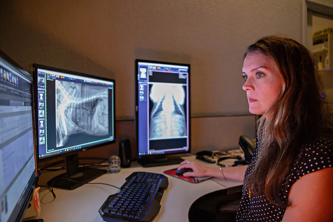
Some dogs do not have any clinical signs of a lung tumor; however, the tumor can be found on chest x- rays. Warning signs of a lung tumor include coughing, which may produce mucous, or blood, loss of appetite, and weight loss. If breathing difficulties are noted in the patient with a lung tumor, diffuse lung disease or infiltration of tumor through the lungs is probable.
The diagnosis of a lung lobe tumor is based on clinical signs, physical examination, chest x-ray findings, and biopsy of the mass. Other tests that may be indicated to help diagnose the problem include bronchoscopy (camera placed down the airways via the mouth), chest CT scan, and thoracoscopy (camera placed through the chest wall).
Treatment.
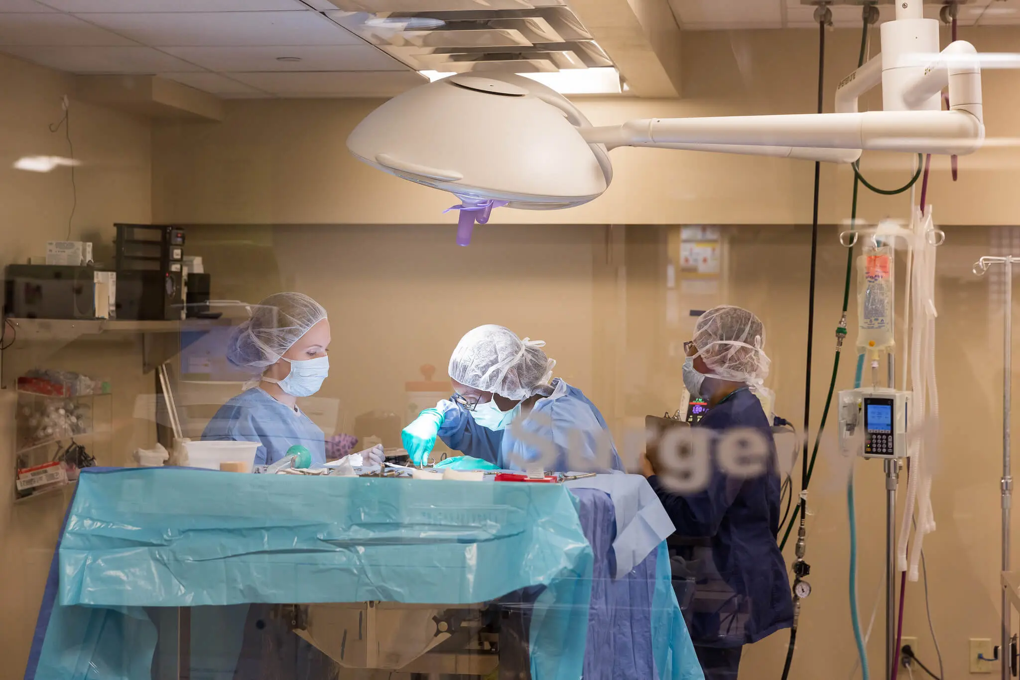
Surgery is recommended to remove primary lung tumors (single tumor) if the patient does not have breathing difficulties. The affected lung lobe is removed via an incision that is made on the side of the  chest. After the lung lobe is removed, a tube is placed through the chest wall and into the chest cavity. The surgical incision is then closed. The chest tube is kept in place to allow for the evacuation of fluid and air from the chest cavity during the first 2-3 days after surgery.
For more information on this subject, speak to the veterinarian who is treating your pet.


