What is upper airway obstruction?
Upper airway obstruction (UAO) in dogs and cats can be a life-threatening emergency as the major source of air into and out of the lungs is partially or completely blocked. The upper airway in this context includes the larynx, pharynx and trachea.
Several mechanical or functional disorders can cause UAO; some of these are due to breed-related anatomy, while others can be caused by an acquired obstructive disease process or the sudden inhalation of a foreign body.
Emergency stabilization is often necessary even before the cause is identified, and definitive treatment may involve surgical, medical, interventional or a combination of treatments.
Clinical signs and symptoms.
The most common sign of a patient with airway obstruction is loud inhaling or exhaling, which can be heard without a stethoscope.
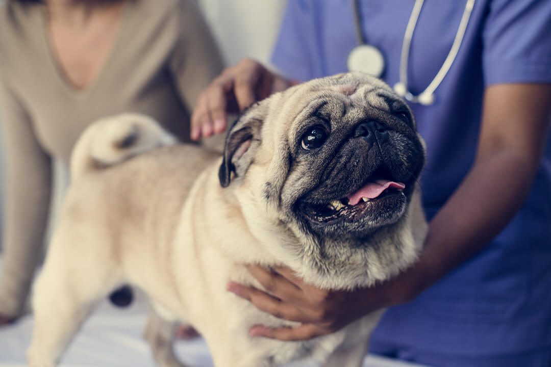
Many owners of breeds with anatomically constricted airways – especially brachycephalic (short-nosed) breeds like English, French or American bulldogs, pugs or Boston terriers – are accustomed to “noisy breathing” or “snoring” from their pets. Unfortunately, these signs can progress with age, exertion, weight gain and in hot or humid weather conditions, and become a true respiratory emergency.
Dogs and cats with near complete or complete airway obstruction often present in a crisis, typically making gasping noises or no noise at all because air is unable to pass.
Respiratory distress can also be seen as attempting specific postures to ease breathing (extending the neck, not laying down), or a blue discoloration to the tongue or gums as oxygenation levels fall to dangerous levels. Because dogs rely on panting as their primary mechanism of cooling, airway obstruction quickly leads to overheating and potential heat stroke.
UAO is less common in cats than dogs, but the signs are similar: Exaggerated, open-mouthed breathing efforts or noisy breathing, especially if the patient is stressed.
Causes.
The most common causes of UAO in dogs are:
- Brachycephalic upper airway syndrome (BUAS)
- Laryngeal paralysis (more common in older, large breed dogs such as Labrador retrievers)
- Tracheal collapse (more common in small breed dogs with progressive tracheal weakness, such as Yorkshire terriers)
Cats are less likely to develop UAO as they are typically indoors and unexposed to heat and exertion. However, brachycephalic cat breeds such as Persians can still develop respiratory distress, especially during travel or stressful veterinary visits.
Both dogs and cats can develop space-occupying structures that obstruct the upper airways. Examples of these are pharyngeal mucoceles (a surgically treatable disease), laryngeal masses or tracheal masses. Because of the progressive growth of these structures, signs usually appear gradually but may be exacerbated by stress and appear suddenly.
Dogs and cats can both inhale foreign bodies that obstruct the larynx or trachea; catching balls that lodge in the back of the throat or a treat that is inhaled rather than swallowed can be life-threatening if not quickly addressed.
Getting a diagnosis.
When a patient is being examined during the emergency intake process, their history, breed and clinical picture are usually very suggestive of UAO and the most likely cause. Although additional testing and examination are helpful in confirming the diagnosis, stabilizing these very fragile patients is the priority, and owners must be prepared to make quick decisions for their pets in emergencies.
The pharynx and larynx can be evaluated with a sedated airway exam by an experienced clinician or, if available, a surgeon. This requires injectable, short-acting anesthesia and evaluation of the size and movement of the structures in the back of the throat. This is typically followed by inserting a breathing tube for a short time until the patient wakes up or, if needed, undergoes surgery.
Visualizing the trachea internally is not always possible without a specialist prepared to perform tracheoscopy (inserting an endoscope into the airway to evaluate for tracheal collapse, masses or foreign bodies). More commonly, the patient’s airways are first evaluated by radiographs (x-rays) either while intubated or once this is safe to perform. Radiographs are also a helpful tool to evaluate the laryngeal structures for the presence of masses. Because they are challenging to interpret, it is always recommended that the images be sent to a radiology specialist.
Treatment.
Stabilization of a patient with UAO is a priority as these patients are often the most emergent in the hospital and present in crisis. Keeping your pet cool and transporting them to the nearest emergency hospital are key. If possible, call the hospital ahead of time so they can prepare for an incoming airway emergency.
Once at the hospital, the emergency team will perform stabilization steps, which include IV access, cooling measures, sedation, oxygen administration, and sometimes the placement of a breathing tube.
If the patient is helped with sedation, oxygen and cooling, and a tentative diagnosis of the type of obstruction has been made, then emergency surgery may be avoidable and the patient may be managed medically.
Surgical procedures to correct the airways of dogs with advanced laryngeal paralysis (laryngeal tie-back procedures) or brachycephalic syndrome (surgery of the nares, soft palate and saccules) are best performed in a proactive and planned fashion rather than as an emergency. Unfortunately, sometimes medical attempts fail and an emergency tracheotomy is necessary for the pet to breathe on their own without a tube, for airway inflammation to decrease or for the pet to have definitive surgery.
In dogs with tracheal collapse, medical management is often recommended to control coughing, distress, inflammation and any possible lower airway infection. If they cannot be stabilized with medical management and short-term intubation, they may need an interventional procedure called tracheal stenting, where a metal stent implant is placed inside the trachea to hold it open.
Upper airway surgical and interventional procedures should be considered and undertaken with care. A risk/benefit analysis should be performed and it’s critical for the patient to have support before, during and after their procedure in a 24/7 facility. Many BluePearl hospitals are equipped with surgery, internal medicine and critical care specialists to manage the many aspects of these cases as a team.
Prevention.
Both American and international veterinary medical associations are taking steps to inform the public of the health and welfare limitations of brachycephalic dogs. Decreasing the demand for these breeds is key to decreasing the morbidity, mortality and owner cost associated with complex respiratory lifesaving care.
Owners of brachycephalic dogs are encouraged to see a veterinary surgeon for airway surgery consultation. This may or may not be necessary and the timing can be best determined by the surgeon. However, being proactive can be lifesaving and prevent potential acute presentations of respiratory distress.
In general, owners of dogs and cats with brachycephalic airway anatomy, tracheal collapse or laryngeal paralysis can make several lifestyle adjustments for their pets:
- Keep pets indoors and air conditioned during warm parts of the day and in humid climates
- Provide them with plenty of cooling areas, shade, rest and fresh water
- Monitor their diet and exercise to help them maintain a lean body weight
- Use sedatives as needed in times of distress or for veterinary visits
- Avoid water activities where immersion is possible
- Use a chest harness instead of a neck collar to prevent pulling on the airways
- Avoid long distance travel if this causes a pet stress
- Avoid putting pets with potential breathing issues in cargo compartments of airplanes
Conclusion.
UAO in dogs and cats can be a stressful experience for owners and dangerous for the pet. Seeking emergency veterinary care if your pet is experiencing signs that are getting worse is key to getting them lifesaving care.
Emergency care or advanced specialty care may be needed for many of these pets.
Airway emergencies can often be avoided by being safe and keeping your pet quiet, calm and cool at home, especially on warm and humid summer days.