Clinicians should take a rational diagnostic approach to episodic weakness in the dog.
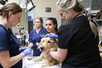
Being faced with a canine patient that has been exhibiting weakness in a paroxysmal manner over a period of several weeks to months can be a challenging diagnostic dilemma for the small animal clinician. As with any complex syndrome, it is best to approach it in a systematic manner.
The cornerstone of any workup must begin with a detailed history from the pet owner including age, breed, sex (neutered or intact), duration of the problem, duration of the episode, is accompanying loss of consciousness (syncope vs. absence seizure), exercise tolerance, cough, polyuria/polydipsia, gastrointestinal disturbance (vomition/diarrhea-melena/hematemesis/ hematochezia), and loss of normal gait or cognition (circling, head pressing, aimless wandering).
After a thorough physical examination, the minimum data base should include: a complete blood count, automated 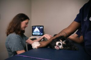 chemistry panel, electrolytes, TT4, thoracic and abdominal radiographs and blood pressure measurement. From the results of these tests, the clinician is able to expand the diagnostic investigation of the patient.
If a cardiac murmur is detected, carefully reviewing the chest x-rays, and pairing those findings with an ECG/echocardiogram, can be diagnostic. If the patient is exhibiting hypertension, a urine protein to creatinine ratio would be valuable in determining if there is a glomerulopathy. Also, if the patient exhibits marked tachycardia, an ECG/abdominal ultrasound may detect a cardiac abnormality or pheochromocytoma.
 Older patients that exhibit profound hypoglycemia should be worked up for a functional islet cell tumor of the pancreas (insulinoma – AIGR, abdominal ultrasound). In dogs whose rear legs collapse and/or exhibit dysphagia, myasthenia gravis should be considered (anti-acetylcholine antibody test, thoracic x-rays – look for thymic mass/megaesophagus).
Bradycardia with hyperkalemia should alert the clinician to possible adrenal insufficiency (ECG,1-hour cortrosyn stimulation test). Anemia in an older large dog may signal the possibility of a bleeding abdominal mass – i.e., splenic hemangiosarcoma (abdominal ultrasound/abdominoacentesis). A profoundly weak dog that has marked hyperglycemia could either have OKA or non-ketotic hyperosmolar encephalopathy.
Young dogs that exhibit vague CNS signs and/or a failure to thrive should be investigated for a portosystemic shunt (serum bile acids, blood NH3, abdominal ultrasound, abdominal CT with dye). In rare instances, dogs with profound hypothyroidism may exhibit profound weakness (FT4, TSH).
Hypercalcemia which may be primary (PTH, ionized Ca++), secondary (dietary), pseudohyyperparathroidism (a paraneoplastic syndrome due to LSA, anal sac carcinoma, multiple myeloma, mammary carcinoma, nasal carcinoma, etc.) can cause weakness and ECG abnormalities (prolonged QT interval, PVCs, etc.). Depending upon the suspected etiology, the following tests may be incorporated (biopsy of mass, serum protein electrophoresis, urine Bence Jones protein analysis). In older patients that exhibit weakness with relatively normal laboratory tests, a possible space-occupying mass should be suspected (detailed CNS exam-cranial nerves, retinal exam, long tract exam, hemi-hopping-followed by CSF tap/MRI).
The above diagnostic approach should give the clinician a template for approaching the patient that exhibits episodic weakness in an orderly manner. The most important two points are (1) Always have an index of suspicion – if one is not open to a possible etiology, a diagnosis will not be made. (2) The value of a detailed history before embarking upon costly diagnostic testing cannot be overemphasized.