Advanced Technology and a Dog Named Maddie
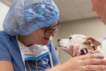
SOUTHFIELD, Mich. – As veterinary medicine gets more and more advanced, veterinarians are increasingly treating pets with sophisticated technology that was originally developed for use in human hospitals.
That technology, and the knowledge of how to use it, recently helped save a lovable Yorkshire terrier named Maddie.
Maddie, who is 7, began breathing heavily and could not get to sleep at all one night while her owners, the Cassel family, were on vacation in Northern Michigan. The family drove the next day to the BluePearl Pet Hospital in Southfield, Mich., cutting their trip short by a day. Maddie had been a heavy breather for years, but this time she was clearly in trouble.
To diagnose and treat Maddie, veterinarians used four different types of medical imaging technology. And Maddie is now breathing better than she has in years.
“To us as a family, it’s like a miracle,” said Maddie’s owner Prudy Cassel, of Flat Rock.
Here is a step-by-step look at how veterinarians used the different types of technology to help Maddie after she came in to BluePearl in mid-July:
- X-rays. Radiographs (X-rays) of Maddie showed she had pneumonia practically filling one lung lobe, which explained part of the reason for her coughing attack. One of BluePearl’s critical care specialists, Ashley Davis, oversaw her intensive care including oxygen therapy, broad spectrum antibiotics, and IV fluids. But the X-rays also showed another concern – a collapsed trachea, which would make it much more difficult for her to take in air normally. Given the severity and location of the collapse, a birth defect leading to progressive obstruction was suspected.
- Ultrasound. Ultrasound is familiar to anyone expecting a baby, but this type of imaging has broader uses in both human and veterinary medicine. BluePearl’s radiologist, Michelle Rose performed an abdominal ultrasound to look for anything else that may have contributed to her sudden symptoms. In addition, she was able to ultrasound Maddie’s trachea to rule out obvious masses or foreign material.
- Bronchoscope. Next, Dr. Jessica Romine, in the Internal Medicine department, used a long thin instrument called a bronchoscope, equipped with a tiny video camera on the end. She inserted this into Maddie’s airway, which proved to Romine the trachea had collapsed. In fact, she could see it was almost completely blocked.
- Fluoroscopy. By this point, Romine knew the best treatment for Maddie would be to place a stent – inserting a woven metal tube to hold the trachea open. But how would she know she had placed a stent of exactly the right size in exactly the right place? She used fluoroscopy, an X-ray-based imaging technique, which allowed her to observe the procedure on a video screen in real time, even as she was performing it. She put two stents in place to provide extra support given the severity of the collapse.
Maddie’s treatment was a team effort, performed by veterinarians with advanced training in emergency, critical care, radiology, and internal medicine.
…but Maddie had a collapsed trachea, which severely inhibited her ability to breathe.
Air normally should pass freely through the trachea on its way to the lungs…
“It’s like a person going in to the hospital,” Cassel. “To know that they can give this kind of care to an animal and be just as successful as on a human being, I think that’s amazing.”
Cassel, who works in billing for a medical practice, said she had no idea that veterinarians have such a wide range of expertise. “Veterinarians have specialties too. Did I know that? No, I didn’t.”
Maddie’s collapsed trachea was a congenital defect, and it explained why she was in such severe respiratory distress. “This pneumonia sent her into a crisis, and it probably wouldn’t have without this congenital defect,” Romine said.
After Romine placed the stents, Maddie was still recovering from pneumonia, but her loud, honking cough had gone away. Even under light anesthesia, Maddie had been coughing in the fluoroscopy procedure, but as soon as the stents were placed she began breathing deeply and quietly.
“She was immediately doing better and we were able to discharge her the next day,’ Romine said. At her first recheck a week later, Maddie’s pneumonia had almost completely resolved. Because she is a young dog, she will need to be monitored over time to ensure the stent remains in the right location, but with medical management, the BluePearl doctors have high expectations for Maddie long-term.
In fact, Cassel says the family had gotten so used to Maddie’s loud breathing, that it took a while to get used to her not doing it. Sometimes she still thinks where’s Maddie? And Maddie is right next to her.
Dr. Romine placed stents, which keep Maddie’s trachea open and have dramatically improved her breathing.