No Need to Dread the Grumpy Limping Cat
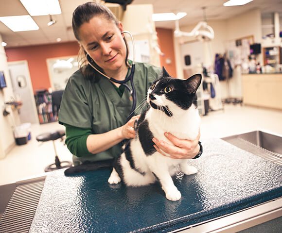
A domestic short-haired cat is on your schedule for evaluation of hind limb lameness and reluctance to jump on furniture. Your first thought may be that you LOVE doing orthopedic examinations on cats, especially the grumpy ones. But then you note the signalment: the cat is 1.5 years old and male neutered. You take a peek through the carrier as he hisses and swats, and you note that he is also overweight. On further questioning, you come to learn that he is an indoor-only cat with no history of trauma.
Eureka! You have instantly narrowed your differentials to spontaneous femoral capital physeal fracture or metaphyseal osteopathy of the femoral neck, and you haven’t even been scratched! You attempt a physical examination where the cat growls throughout but seems especially displeased with bilateral hip extension. Without further hesitation, you recommend a touch of sedation and radiographs.
Evaluating Non-Traumatic Fractures
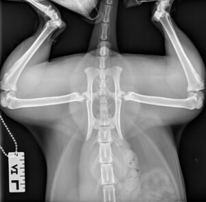
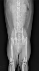
 After a lovely intramuscular sedation cocktail, you are able to evaluate the cat and detect crepitus and decreased range of motion in both coxofemoral joints. The remainder of your orthopedic examination is unremarkable. A ventrodorsal radiograph of the pelvis, as well as a frog-legged view were obtained and revealed lysis and bony remodeling of both femoral necks and a pathologic fracture of the capital physis on the right. You diagnose metaphyseal osteopathy with capital physeal fracture.
Once you review the radiographs with the owner, the owner is skeptical as to how this could have occurred with no trauma history. There are two main non-traumatic conditions of the femoral head and neck in cats, the first being spontaneous capital physeal fractures and the second being metaphyseal osteopathy.
Diagnosing and Treating Spontaneous Capital Physeal Fractures and Metaphyseal Osteopathy
Spontaneous capital physeal fractures usually occur in young, overweight neutered male cats. Clinical signs can vary from acute or chronic lameness. Cats are often reluctant to jump. Physical examination reveals decreased range of motion, pain and crepitus on hip extension. The diagnosis is best made on radiographs, specifically the frog-legged view, which enables one to clearly see the displacement of the epiphysis from the metaphysis.
Risk factors for developing these spontaneous capital physeal fractures in cats are:
- Male
- Neutered
- Delayed physeal closure
- Overweight
 Cats with metaphyseal osteopathy have a very similar presentation; however, radiographically there is severe lysis and bony remodeling at the femoral neck. These can also be associated with pathologic fractures through the capital physis or femoral neck. These fractures are unlikely to heal with surgical stabilization because of the remodeling and compromised vasculature. Resorption is thought to occur in the femoral neck, which then leads to the epiphyseal separation.
In most cases of metaphyseal osteopathy and spontaneous capital physeal fractures, a femoral head and neck ostectomy is recommended. The majority of cats recover well and are reported to regain normal use of the affected limb.
To discuss one of these cases, or any other surgical case, find a BluePearl Pet Hospital near you.


